![]() Figure 5 of
Kompella, Mol Vis 2006;
12:1185-1198.
Figure 5 of
Kompella, Mol Vis 2006;
12:1185-1198.
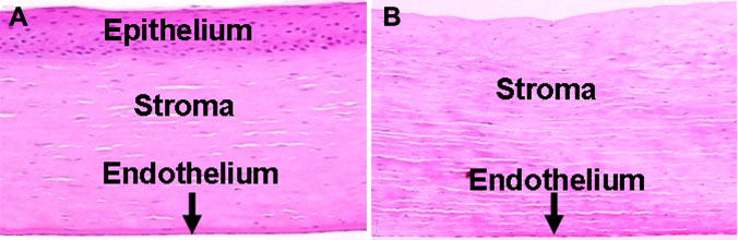
Figure 5.
Corneal epithelium is a substantial barrier for nanoparticle delivery in the bovine ex vivo eye model. After instillation of a 50 μl drop of 10 mg/ml plain nanoparticles and 5 or 60 min uptake study at 37 °C, the tissue layers were isolated, homogenized, and particle uptake was quantified. The figure shows bovine cornea with (A) and without epithelium (B) and uptake of nanoparticles at 5 min (C, D) and 60 min (E, F) in intact bovine eyes (C, E) or those devoid of corneal epithelium (D, F). The data are expressed as mean±SD for n=3. Magnification in A and B was 5X. The asterisk indicates a p<0.05 compared to the group on the left with epithelium. The double asterisk indicates a p<0.05 compared to stroma, endothelium, and aqueous humor.