![]() Figure 4 of
Kompella, Mol Vis 2006;
12:1185-1198.
Figure 4 of
Kompella, Mol Vis 2006;
12:1185-1198.
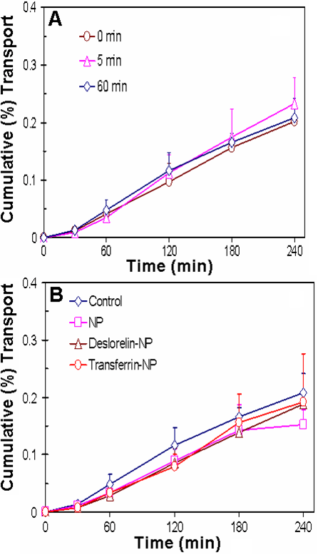
Figure 4.
Corneas retain barrier integrity during the ex vivo bovine eye study with or without nanoparticle treatment. A 50 μl drop of either assay buffer (control) or 1 mg/ml nanoparticle suspension was topically instilled in the ex vivo bovine eye model maintained at 37 °C. After 0, 5, and 60 min of exposure to assay buffer and 60 min exposure to nanoparticle suspensions, corneas were isolated and permeability of 3H-mannitol, a paracellular marker, was assessed. Percent cumulative transport is plotted for corneas obtained after assay buffer drop administration and ex vivo study termination at various time points (A) or after nanoparticle suspension administration followed by ex vivo study termination at 60 min (B). The data are expressed as mean±SD for n=4.