![]() Figure 2 of
Kompella, Mol Vis 2006;
12:1185-1198.
Figure 2 of
Kompella, Mol Vis 2006;
12:1185-1198.
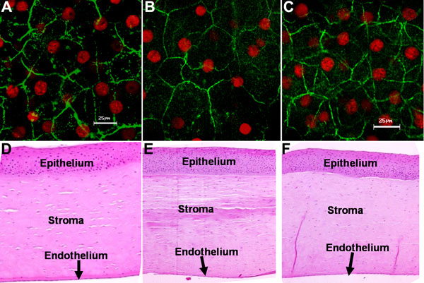
Figure 2.
Corneal epithelial tight junctional architecture and histology in a bovine ex vivo eye model treated with plain buffer. A, B, and C represent confocal images of bovine corneas double labeled with anti-ZO1 antibody (green) and propidium iodide (red) at 0 (control), 5, and 60 min, respectively, in the ex vivo study with plain buffer treatment at 37 °C. D, E, and F show representative histology pictures (magnification 5X) of cornea after hematoxylin and eosin staining at 0 (control), 5, and 60 min, respectively, in the ex vivo study with plain buffer. The confocal images were obtained at a magnification of 63X.