![]() Figure 10 of
Kompella, Mol Vis 2006;
12:1185-1198.
Figure 10 of
Kompella, Mol Vis 2006;
12:1185-1198.
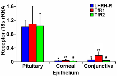
Figure 10.
Differential expression of LHRH and transferrin receptors on various bovine ocular tissues. The fold difference (2-ΔCt) of LHRH receptor (blue bars), transferrin receptor 1 (red bars) and transferrin receptor 2 (green bars) between the corneal epithelium and conjunctiva is presented as mean±SD for n=3 samples. The asterisk indicates a p<0.05 compared with LHRH-R, the double asterisk indicates a p<0.05 compared with TfR1, and the sharp (hash mark) indicates a p<0.05 compared with TfR2, in the pituitary.