![]() Figure 6 of
Ryan, Mol Vis 2006;
12:1175-1184.
Figure 6 of
Ryan, Mol Vis 2006;
12:1175-1184.
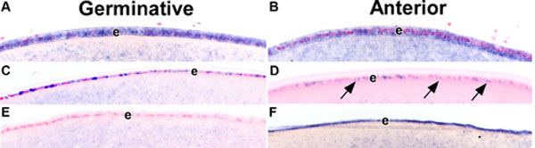
Figure 6.
MicroRNAs reveal distinct expression patterns in the lens epithelium. Neonatal (A and B) and adult (C-F) mouse lens were processed for in situ hybridization with digoxygenin-labeled antisense probes for mir-184 (A-D) -204 (F) and a scrambled oligonucleotide sense (E) control. Uniform expression of mir-184 is seen in epithelial (e) cells from the germinative (A) and anterior (B) regions of the lens. In contrast, mir-184 expression is more uniform in the epithelial (e) cells of the germinative (C) compared with the anterior (D; arrows) regions. Expression of mir-204 (F) is uniformly detected in the epithelial cells of the anterior region of the lens. Staining detected in the region beneath the epithelial layer with the sense control (E), is believed to be non-specific.