![]() Figure 4 of
Ryan, Mol Vis 2006;
12:1175-1184.
Figure 4 of
Ryan, Mol Vis 2006;
12:1175-1184.
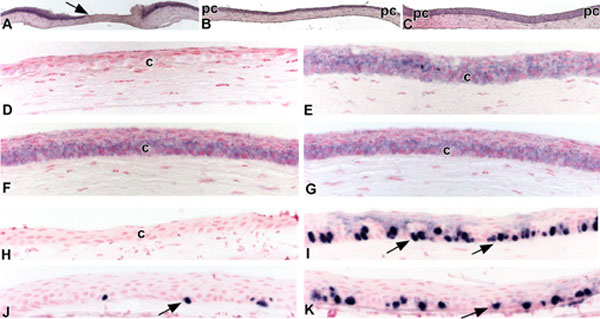
Figure 4.
Mir-184 expression in mouse corneal epithelium is independent of proliferation. A-C: Application of a rotating diamond burr to the surface of the central cornea resulted in the removal of the corneal epithelium (A; arrow), whereas the peripheral corneal epithelium was intact (pc). Within 24 h (B) the corneal epithelial surface was reepithelialized, and by 48 h (C) the corneal epithelium returned towards a normal phenotype. D-G: Corneal tissues were processed for in situ hybridization with a digoxygenin-labeled antisense probe for mir-184. Twenty-four h post wounding, no signal for mir-184 was detected in the re-epithelialized central corneal epithelium (D); however, a strong signal was observed throughout the adjacent (pc) nonwounded corneal epithelium (E). Forty-eight h post wounding, strong expression of mir-184 is seen in both the reepithelialized (F) and adjacent (pc) nonwounded (G) corneal epithelia. H-K: Twenty-four (H,I) and 48 h (J,K) following the creation of scrape wounds, mice received a pulse of BrdU interperitoneally to label those cells in the S phase of division. Mice were sacrificed 1 h later. Twenty-four h postwounding, no BrdU labeled cells were detected in the reepithelialized central corneal epithelium (H); however, numerous BrdU labeled basal cells (arrows) were noted in the adjacent (pc) nonwounded corneal epithelium (I). Forty-eight h post wounding, some BrdU labeled basal cells (arrows) were noted in the reepithelialized corneal epithelium (J), while there was a reduction in BrdU-labeled basal cells (arrows) in the adjacent (pc) nonwounded (K) corneal epithelium.