![]() Figure 2 of
Ryan, Mol Vis 2006;
12:1175-1184.
Figure 2 of
Ryan, Mol Vis 2006;
12:1175-1184.
Figure 2.
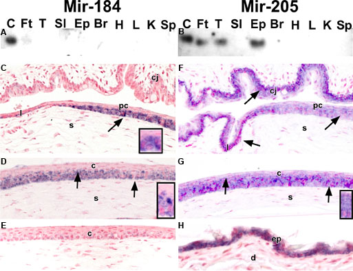
Epithelial miRNAs exhibit tissue and cell-type specificity. A and B: Total RNA (10 μg) from mouse corneal epithelium (C), footpad epithelium (Ft), tongue (T), small intestine (SI), epidermis (Ep), brain (Br), heart (H), liver (L), kidney (K), and spleen (Sp) were analyzed by northern hybridization with 32P-labeled oligonucleotides to mir-184 (A) and -205 (B). Mir-184 was abundant solely in corneal epithelium (A), whereas mir-205 was present in the RNA from several epithelial tissues (B). C-H: Mouse anterior segmental epithelia (C-G) and epidermis (H) were processed for in situ hybridization with digoxygenin-labeled antisense probes for mir-184 (C and D) and mir-205 (F-H) and a scrambled oligonucleotide sense (E) control. Mir-184 is expressed in corneal (c) but not limbal (l) and conjunctival (cj) epithelia. Mir-184 RNA is observed (arrows) in basal and immediately suprabasal cells of the corneal epithelium. Little if any signal for mir-184 is seen in the wing and superficial cells of the corneal epithelium (C and D) Boxed areas in C and D show mir-184 expression restricted to basal and wing cells. Mir-205 RNA is present in all layers of the peripheral (pc) and central corneal (c) epithelia (F and G) as well as the limbal (l) and conjunctival (l) epithelia (F). Boxed area in G shows mir-205 expression in all layers of the corneal epithelium. A strong signal for mir-205 is also detected throughout the mouse epidermis (e; H). In the image, s denotes the stroma and d indicates dermis.