![]() Figure 3 of
Jin, Mol Vis 2006;
12:1167-1174.
Figure 3 of
Jin, Mol Vis 2006;
12:1167-1174.
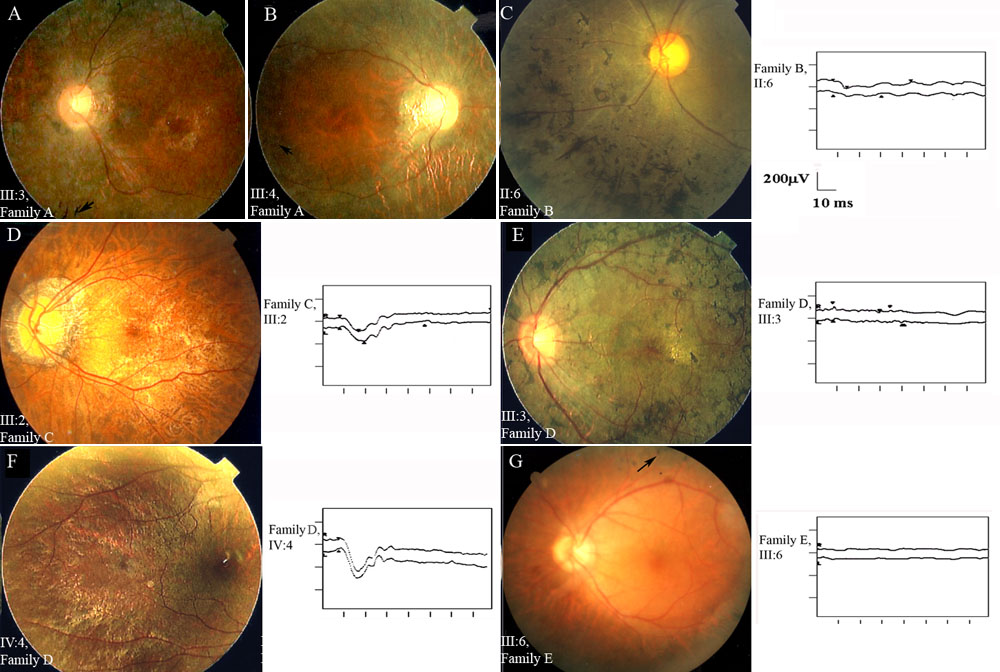
Figure 3. Representative fundus photographs and ERG results of the XLRP individuals
A: Family A, patient III:3: Fundus photograph showing bone spicule-like pigmentation (arrowhead) and macular degeneration. B: Family A, patient III:4: Fundus photograph showing leopard fundus and a few pigmentary changes (arrowhead) in the midperipheral retina. C: Family B, patient II:6: Fundus photograph showing extensive bone spicular changes and the maximal combined response (right). D: Family C, patient III:2: Fundus photograph from a carrier showing extensive myopic chorioretinal degeneration with peripapillary atrophy (left) and the maximal combined response (right). E: Family D, patient III:3: Fundus photograph showing the accumulation of bone spicules in the midperiphral fundus (left) and the maximal combined response (right). F: Family D, patient IV:4: Fundus photography of a carrier showing tapetal-like reflex (left) and maximal combined response (right). G: Family E, patient III:6: Fundus photography showing bone spicular pigmentations in midperipheral fundus (left) and maximal combined response (right).