![]() Figure 1 of
Sakamoto, Mol Vis 2006;
12:117-124.
Figure 1 of
Sakamoto, Mol Vis 2006;
12:117-124.
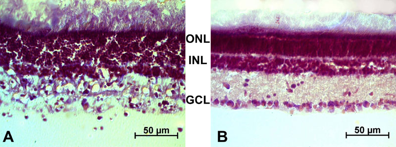
Figure 1. Effect of kainic acid treatment on the retina: histological evaluation of kainic acid treatment
Retinas were dissected 48 h after intraocular injection of 200 nmol kainic acid (KA; A) or saline (control; B), sectioned, and stained with cresyl violet. Micrographs show that in the KA-treated retina, most of the inner nuclear layer (INL) has degenerated. No significant alteration was apparent in the outer nuclear layer (ONL). A few ganglion cells were still visible in the ganglion cell layer (GCL). The scale bars represent 50 μm.