![]() Figure 2 of
Perra, Mol Vis 2006;
12:1136-1142.
Figure 2 of
Perra, Mol Vis 2006;
12:1136-1142.
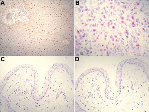
Figure 2.
Immunohistochemical study of control sections. A: Rat liver. B: Human melanoma. C and D: Normal conjunctiva. A and C: 8-OHdG staining. B and D: p53 staining. Numerous 8-OHdG positive hepatocytes were observed (A). p53 nuclear staining was evident in scattered tumor cells (B). No immunostaining for 8-OHdG and p53 in normal conjunctiva was observed (C and D). The original magnification for A, C, and D was 200x and the original magnification for B was 400x.