![]() Figure 5 of
Chudgar, Mol Vis 2006;
12:1117-1126.
Figure 5 of
Chudgar, Mol Vis 2006;
12:1117-1126.
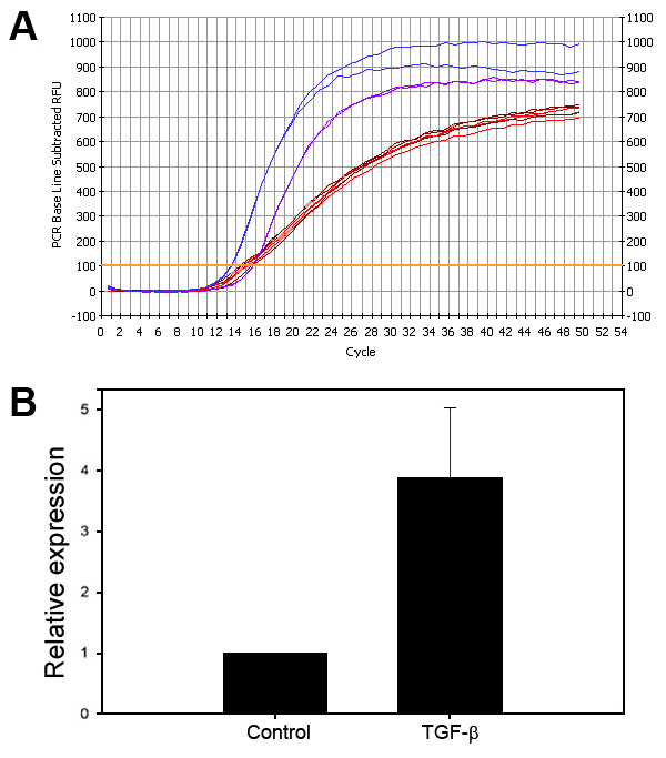
Figure 5.
Quantification of the relative changes in the levels of CTGF mRNA in HTM cells treated with TGF-β1. To determine the relative difference in CTGF expression induced by TGF-β1 in HTM cells, confluent cultures of HTM cells were serum starved for 24 h and treated with 20 ng/ml TGF-β1 for 4 h. Total RNA extracted from 5 sets of control and TGF-β1-treated samples was subjected to real-time quantitative RT-PCR analysis. A: Logarithmic fluorescence history versus cycle number of CTGF from control and TGF-β1-treated HTM cells and housekeeping β-actin gene. The trace colors indicate: CTGF in TGF-β-treated HTM cells (blue); CTGF in control HTM cells (purple); β-actin in TGF-β1-treated cells (brown); and β-actin in control cells (red). A representative data of two individual samples is shown. Real-time quantification of CTGF was normalized to the threshold cycle (CT) value of β-actin where CT equals the PCR cycle number at which the amount of amplified product reached 100 relative fluorescence units (RFU). B: Relative fold increase in CTGF expression with TGF-β treatment in HTM cells. Based on the mean values of five individual samples, the increase in CTGF expression with TGF-β1 treatment was found to be 3.89 fold.