![]() Figure 7 of
Takada, Mol Vis 2006;
12:1108-1116.
Figure 7 of
Takada, Mol Vis 2006;
12:1108-1116.
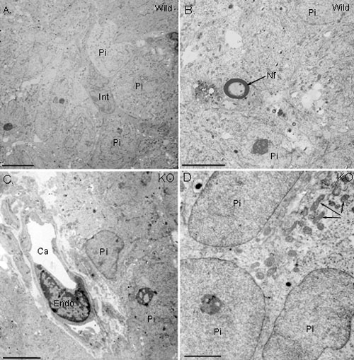
Figure 7. Ultrastructure of WT and RS1-/Y mouse pineal
Electron micrographs showing parts of the superficial pineal gland of the WT mouse (A,B) and RS-KO mouse (C,D). Morphology of the knockout mouse pineal is not different from the wild-type mouse. "Int" represents interstitial cell, "Pi" represents pinealocyte, "Endo" represents endothelial cell, "Ca" represents capillary lumen, "Nf" represents myelinated nerve fiber, arrows in Panel D indicate mitochondria. The scale bar represents 5 μm in A-C and 3 μm in D.