![]() Figure 3 of
Takada, Mol Vis 2006;
12:1108-1116.
Figure 3 of
Takada, Mol Vis 2006;
12:1108-1116.
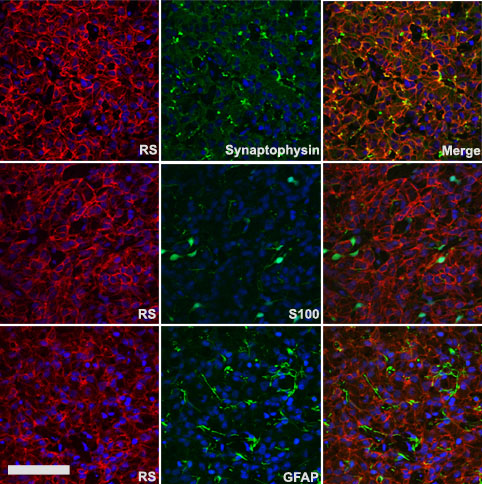
Figure 3. Colocalization of retinoschisin with synaptophysin, S-100, and GFAP in the rat pineal
Immunofluorescence double labeling in the rat pineal with antibodies against retinoschisin (red), synaptophysin (green), S-100 (green), and GFAP (green). A membranous pattern of staining with retinoschisin antibody colocalized with synaptophysin staining in pinealocytes and their synaptic vesicles, but not with S-100 and GFAP in interstitial glial cells. The scale bar represents 75 μm.