![]() Figure 1 of
Nallathambi, Mol Vis 2006;
12:1086-1092.
Figure 1 of
Nallathambi, Mol Vis 2006;
12:1086-1092.
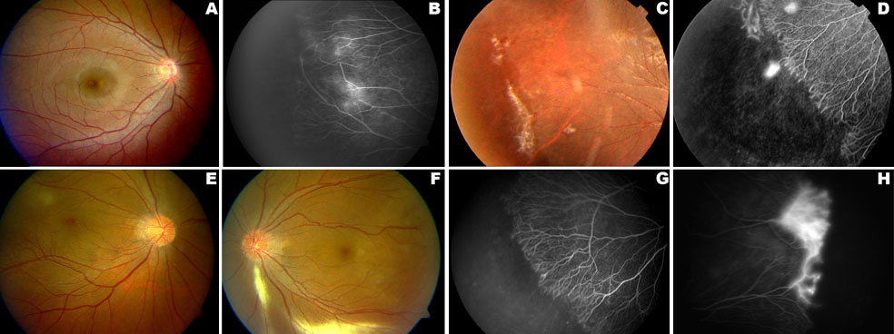
Figure 1. Fundus photography and fluorescein angiography of the FEVR patients with FZD4 gene mutations
A,B: The fundus picture of patient IV: 3 from family one, which shows macular ectopia of the right eye; the angiogram demonstrates the peripheral avascular zone. C,D: In the second family, ophthalmic examination of a patient (III: 7) shows the peripheral avascular zone with fibrous proliferation in the right eye; angiogram reveals neovascularization with peripheral avascular zone. E-H: Ophthalmoscopic appearance of the patient (II: 1) from third family, demonstrates macular ectopia in both eyes with medullated nerve fibers in the left eye. The angiogram shows the peripheral avascular zones in both eyes with leaking fibrovascular proliferation in the left eye.