![]() Figure 3 of
Colitz, Mol Vis 2006;
12:1067-1076.
Figure 3 of
Colitz, Mol Vis 2006;
12:1067-1076.
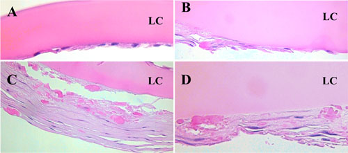
Figure 3.
Photomicrographs of 4 canine anterior capsulotomy samples from patients with naturally occurring cataracts. A,B: The flattened morphology of cataractous LEC. In B, the LEC are beginning to become multilayered. C,D: The extensive multilayered LEC that are clinically apparent as subcapsular plaques. LC=anterior lens capsule; hematoxylin and eosin stain; magnification 400x.