![]() Figure 2 of
Colitz, Mol Vis 2006;
12:1067-1076.
Figure 2 of
Colitz, Mol Vis 2006;
12:1067-1076.
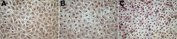
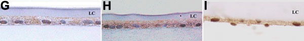
Figure 2.
Immunocytochemical staining for TERT and bar graph representation of immunostaining results in the central (A,D,G), germinative (B,E,H), and equatorial (C,F,I) regions of anterior lens capsule whole mounts from normal canine lenses. Magnification 400x. Central LEC (A,D) had significantly more cytoplasmic staining than combined nuclear and cytoplasmic staining (p<0.001). Germinative LEC (B,E) had equal numbers of cells with cytoplasmic and nuclear plus cytoplasmic staining (p>0.05). Equatorial LEC (C,F) had significantly more cytoplasmic plus nuclear staining than exclusively cytoplasmic staining (p<0.001). Immunohistochemical staining of whole lenses (G,H,I) demonstrated immunopositivity that was consistent with whole mount samples.