![]() Figure 5 of
Gendron, Mol Vis 2006;
12:108-116.
Figure 5 of
Gendron, Mol Vis 2006;
12:108-116.
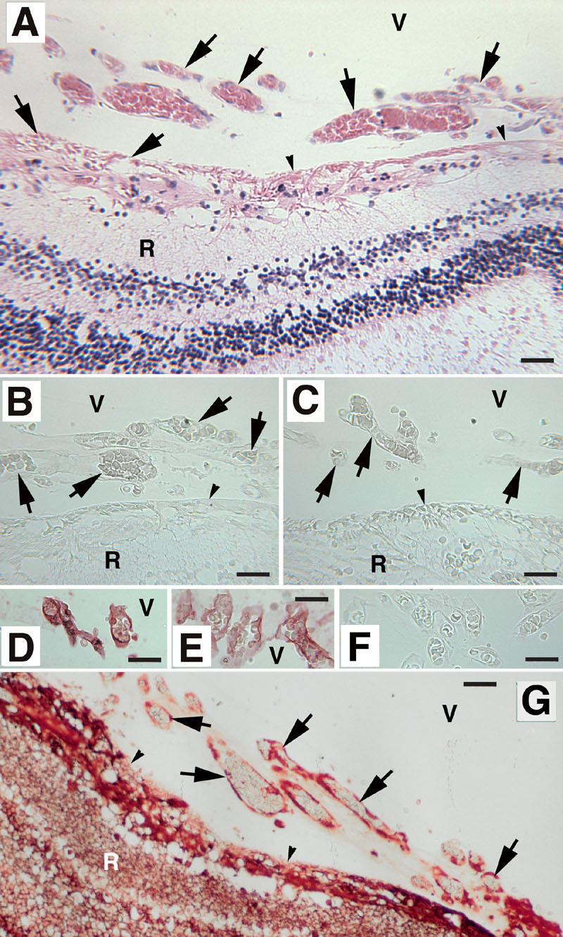
Figure 5. Analysis of Tbdn-1 protein expression in fetal human eye and in a case of human retinopathy of prematurity
A-C,F,G: Retinopathy of prematurity. D,E: Normal fetal human eye. Panels B-F were stained for Tbdn-1 expression. The small proliferative vitreal-retinal vessels (arrows in H&E stained low power section shown in A) growing from the retina into the vitreous humor in the ROP sample show little to no Tbdn-1 expression (B,C). F: ROP sections incubated with preimmune IgY showed no staining. Tbdn-1 expressed by blood vessels within Cloquet's Canal (D) and tunica vasculosa lentis (E) in a 19-week normal fetal human eye specimen without ROP. Blood vessels stain intensely red with antibody to Tbdn-1. G: Expression of α-tubulin in the ROP specimen showing high levels of the ubiquitous α-tubulin protein staining (red) both in the endothelial cells of the diseased neovascular areas of the vitreal-retinal regions and in the nonpathological areas of the ROP retina. A-C,G: The vitreous body is oriented at the top of the panel. The vitreous (V) and retina (R) are identified. Scale bars represent 25 μm. The retinal inner limiting membrane is indicated by small arrowheads. Original magnification was 250x. No counterstain was used in micrographs B-G in order to be able to detect any low levels of Tbdn staining reactions (B-F) and in order to display the high level of ubiquitous tubulin staining in the ROP specimens (G). Micrographs shown are representative experiments.