![]() Figure 1 of
Gendron, Mol Vis 2006;
12:108-116.
Figure 1 of
Gendron, Mol Vis 2006;
12:108-116.
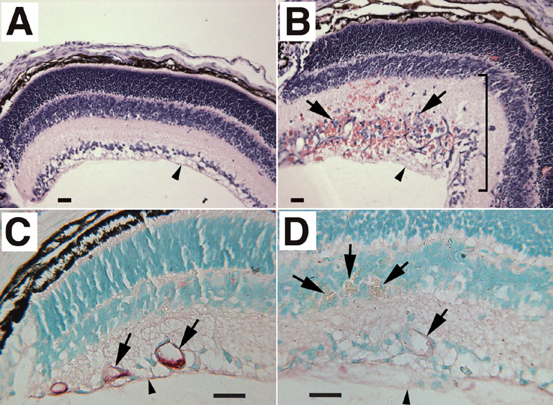
Figure 1. Analysis of Tbdn-1 expression in oxygen-induced retinopathy in mice
A: Control normoxia-reared mice do not show any retinal pathology. B: In striking contrast, hyperoxia-reared mice present retinal lesions displaying retinal neovascularization, fibrovascular growth, and large areas with blood vessel leakage. Arrows point to retinal blood vessels, and the bracket indicates a large area of retinal lesion. Tbdn-1 immunohistochemical analysis of retinal vessels in sections of control normoxia-reared (C) show robust Tbdn expression in controls (red staining seen in vessels marked with arrows) and almost complete loss of retinal blood vessel Tbdn expression in hyperoxia-reared (D) neonatal mice (little to no red staining seen in vessels indicated with arrows). In all panels, day of analysis is P17 and vitreous body is oriented at bottom of panel. Scale bars represent 25 μm. The retinal inner limiting membrane is indicated by small arrowheads. Original magnification was 100x in A,B and 250x in C,D. Counterstain in panels A,B is hematoxylin and eosin. Counterstain in panels C,D is methyl green. Micrographs shown are representative experiments.