![]() Figure 6 of
Johnson, Mol Vis 2006;
12:1057-1066.
Figure 6 of
Johnson, Mol Vis 2006;
12:1057-1066.
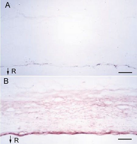
Figure 6.
Immunostaining of human sclera for PRELP. Immunohistochemical staining of frozen sclera obtained from the anterior region of a 36-year-old male donor with anti-PRELP using the Vectastain ABC kit. A: The negative control, where non-immune rabbit serum was substituted for anti-PRELP in the primary antibody incubation. B: Positive staining using the anti-PRELP antibody. The top of the slide represents the outer sclera. The bottom portion of the slide represents the inner, retinal side of the sclera (R). The scale bars represent 150 μm.