![]() Figure 4 of
Ohno-Matsui, Mol Vis 2006;
12:1022-1032.
Figure 4 of
Ohno-Matsui, Mol Vis 2006;
12:1022-1032.
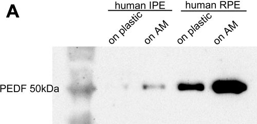
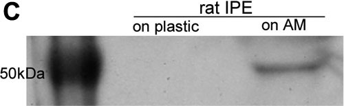
Figure 4.
Increased PEDF protein secretion by IPE cells cultured on AMs. Cells were conditioned in serum free medium for 48 h before harvesting to determine the protein concentration. Equal amounts of secreted protein (8 μg) were applied to each lane. A: Representative western blot analysis of PEDF protein using supernatants from human IPE and RPE cells. PEDF protein levels are increased by culturing cells on AMs both for human IPE cells (lanes 1 and 2) and RPE cells (lanes 3 and 4). The PEDF protein levels are higher in human RPE cells than in IPE cells. B: Quantitative analysis of proteins confirmed that the level of PEDF in human IPE or RPE cells cultured on AMs is higher than that in cells on plastic dish. The value shown is the ratio of the density of PEDF signal to that of IPE cells cultured on AM. PEDF secreted by IPE cells is lower than RPE cells. The asterisk indicates a significant difference (p<0.05, Mann-Whitney analysis). C: Representative western blot analysis of PEDF protein using supernatants from rat IPE cells demonstrating a marked increase in PEDF protein secretion when cells are cultivated on AMs.