![]() Figure 3 of
Ohno-Matsui, Mol Vis 2006;
12:1022-1032.
Figure 3 of
Ohno-Matsui, Mol Vis 2006;
12:1022-1032.
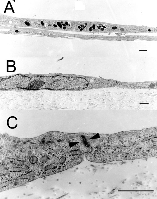
Figure 3.
Transmission electron micrographs of rat IPE cells cultured on uncoated plastic dish (A) or on denuded AMs (B,C) for 14 days. Electron micrographs show a tight monolayer of IPE cells (A) with junctional specializations (C, arrowheads) growing over AM, which is different from the multilayer appearance of cells cultured on plastic dish (A). The scale bars represent 1 μm.