![]() Figure 2 of
Ohno-Matsui, Mol Vis 2006;
12:1022-1032.
Figure 2 of
Ohno-Matsui, Mol Vis 2006;
12:1022-1032.
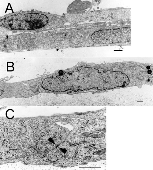
Figure 2.
Representative photographs of transmission electron micrographs of human IPE cells cultivated on uncoated plastic dish (A) or on AMs (B,C) for 14 days. Electron micrographs show a tight monolayer of IPE cells growing over the AM (B), while IPE cells cultured on plastic dish are elongated and multilayered (A). Electron micrograph shows junctional specialization (arrowhead) between adjacent cells cultured on AMs (C). The scale bars represent 1 μm.