![]() Figure 1 of
Ohno-Matsui, Mol Vis 2006;
12:1022-1032.
Figure 1 of
Ohno-Matsui, Mol Vis 2006;
12:1022-1032.
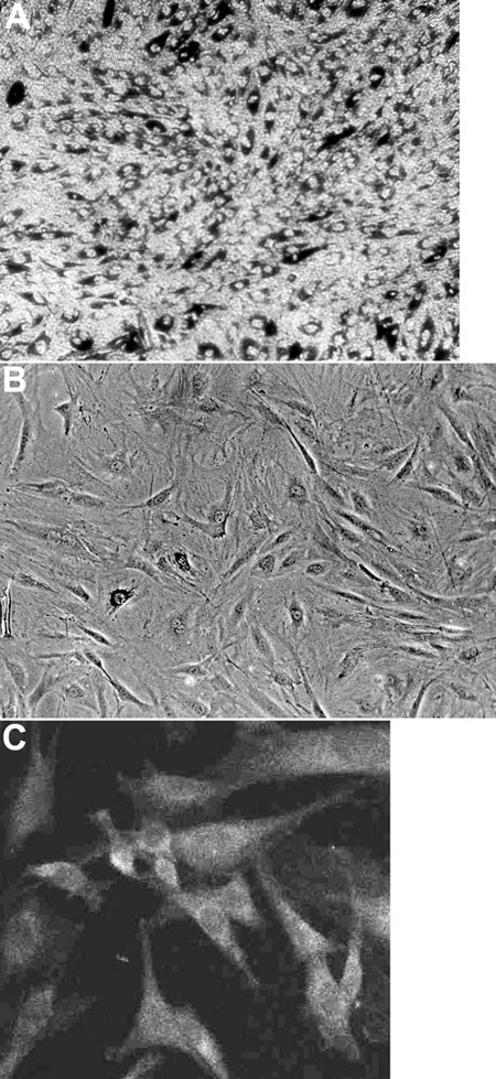
Figure 1.
Photomicrographs of newly-isolated rat IPE cells in culture (A) and at passage 2 (B). The cells are pigmented as viewed through a phase contrast filter (C). The cells are positively labeled with anti-cytokeratin, pan-antibody, indicating their epithelial origin.