![]() Figure 6 of
Qin, Mol Vis 2006;
12:1001-1008.
Figure 6 of
Qin, Mol Vis 2006;
12:1001-1008.
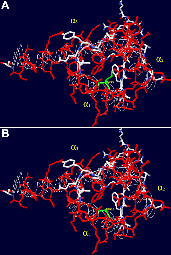
Figure 6.
View of the crystal structure of the homeodomain in PAX3. Three α-helixes are shown in red. The Leu (A) to Pro (B) mutation in helix 1 is shown in green. The sixteenth Leu residue in helix 1 is important in controlling how helix 1 packs against helix 3, stabilizing the folded structure. The program Swiss-PdbViewer was used [37].
