![]() Figure 2 of
Chen, Mol Vis 2006;
12:983-994.
Figure 2 of
Chen, Mol Vis 2006;
12:983-994.
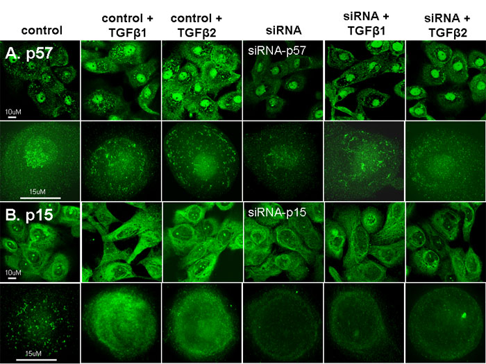
Figure 2.
Immunofluorescent staining, LSCM (first row) and deconvolution microscopy (second row) for A: p57 in control, siRNA-p57 treated before and following TGF-β1 or TGF-β2 treated (1 ng/ml) in primary cultured human limbal epithelial cells. B: p15 in control, siRNA-p57 treated before and following TGF-β1 or TGF-β2 treated in primary cultured human limbal epithelial cells.