![]() Figure 2 of
Mader, Mol Vis 2006;
12:915-930.
Figure 2 of
Mader, Mol Vis 2006;
12:915-930.
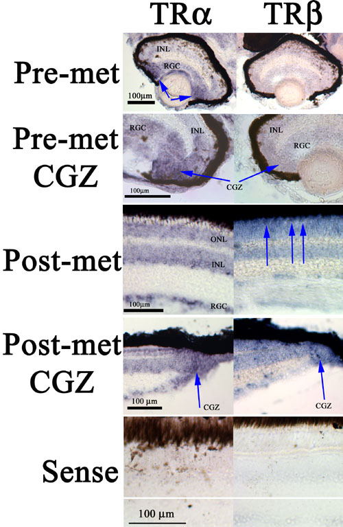
Figure 2. Patterns of TRα and TRβ expression in pre- and postmetamorphic flounder retina
Non-isotopic in situ hybridization analysis revealed evidence for the expression of TRα (left column) and TRβ (right column) in pre- and postmetamorphic retinas as indicated; sense probe controls for each target are presented at the bottom of the figure. TRα expression is particularly strong at the circumferential germinal zone (CGZ; arrows), a region of substantial cytogenesis. In postmetamorphic retinas TRα expression displays "hot spots" (arrows) at the distal outer nuclear layer (ONL), corresponding to the location of cone photoreceptors [32], and also at the CGZ. INL represents inner nuclear layer and RGC represents retinal ganglion cell layer.