![]() Figure 8 of
Steiner-Champliaud, Mol Vis 2006;
12:892-901.
Figure 8 of
Steiner-Champliaud, Mol Vis 2006;
12:892-901.
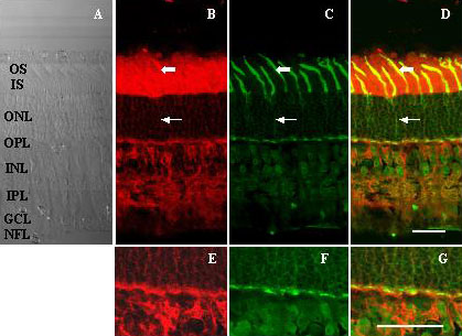
Figure 8. Double staining for Rs1 and PNA gives partial overlap
Adult pig retinal sections (A) stained for Rs1 (B) or PNA lectin (C) showed correspondence of label especially at the level of the cone matrix sheaths (thick arrows), as well as surrounding cell bodies within the ONL (thin arrows). Although the two stains were prominent in the OPL and INL, they did not precisely correspond, with Rs1 localizing more to post-synaptic elements while PNA bound to presynaptic sites (E-G). Scale bar in D represents 30 μm for Panels A-D, and scale bar in G represents 30 μm for Panels E-G. The inner segments (IS), outer segments (OS), outer nuclear layer (ONL), the outer plexiform layer (OPL), inner nuclear layer (INL), inner plexiform layer (IPL), ganglion cell layer (GCL), and nerve fiber layer (NFL) are identified.