![]() Figure 6 of
Steiner-Champliaud, Mol Vis 2006;
12:892-901.
Figure 6 of
Steiner-Champliaud, Mol Vis 2006;
12:892-901.
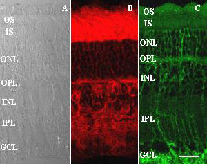
Figure 6. Immunohistochemistry of Rs1 and β2 laminin shows partial overlap
As before, incubation of adult pig retinal sections (A) with Rs1 antibody (B) gave strong staining within the interphotoreceptor matrix, OPL, INL, and IPL, as well as outlining cell bodies in the ONL. We were unable to perform double label immunohistochemistry with the D5 anti-β2 laminin antibody, which works exclusively on unfixed tissue, but the distribution pattern for this antibody partly overlapped with that of Rs1, with label surrounding cell bodies in the ONL and INL, binding to the OPL, and following the trajectories of radial Müller glia and their endfeet (C). Scale bar in C represents 30 μm for all panels. The inner segments (IS), outer segments (OS), outer nuclear layer (ONL), the outer plexiform layer (OPL), inner nuclear layer (INL), inner plexiform layer (IPL), ganglion cell layer (GCL), and nerve fiber layer (NFL) are identified.