![]() Figure 4 of
Steiner-Champliaud, Mol Vis 2006;
12:892-901.
Figure 4 of
Steiner-Champliaud, Mol Vis 2006;
12:892-901.
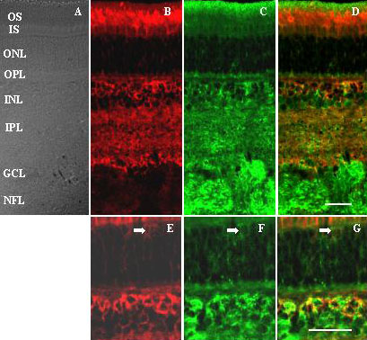
Figure 4. Double label immunohistochemistry of Rs1 and αB crystallin in adult pig retina
Frozen sections of retina (A) were co-incubated with antibodies to Rs1 (B,E) and αB crystallin (C,F). Staining for both antibodies was strong in the interphotoreceptor matrix and IS and OS, as well as in the INL and IPL. In addition αB crystallin immunoreactivity was intense overlying the nerve fiber fundles in the NFL. Staining for both proteins was moderate within the ONL, outlining cell bodies (E,F). Overlay of the two images (D,G) showed label co-segregated within the proximal interphotoreceptor matrix, throughout the INL and IPL, and around cells in the ONL (arrows, E-G). Scale bar in D represents 30 μm for Panels A-D, scale bar in G represents 30 μm for Panels E-G. The inner segments (IS), outer segments (OS), outer nuclear layer (ONL), the outer plexiform layer (OPL), inner nuclear layer (INL), inner plexiform layer (IPL), ganglion cell layer (GCL), and nerve fiber layer (NFL) are identified.