![]() Figure 2 of
Steiner-Champliaud, Mol Vis 2006;
12:892-901.
Figure 2 of
Steiner-Champliaud, Mol Vis 2006;
12:892-901.
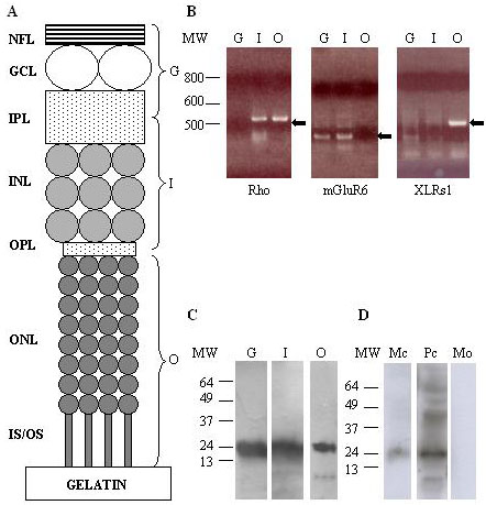
Figure 2. Vibratome separation of retinal layers and RT-PCR and western blot analyses of Rs1
A: In schematic representation, adult mouse retinas were placed photoreceptor-side down on a gelatin block and sectioned at three levels to separate ganglion cell layer (G), inner nuclear layer (I) and outer nuclear layer (O). B: These samples were analyzed by RT-PCR for the presence of specific markers and Rs1 mRNA. Amplification of rhodopsin-specific primers (Rho) detected transcripts in O as expected, but also in I, indicating contamination of the latter with photoreceptor material. There were no rhodopsin transcripts in G. Amplification of mGluR6-specific primers revealed product in I as expected, and also in G from inner plexiform layer (IPL). There was no signal in O. Rs1 transcripts were only detectable in O. C: Anti-Rs1 western blotting of the three samples showed dense bands about 25 kDa in each fraction. D: Anti-Rs1 western blotting of conditioned medium from adult pig photoreceptor cultures (Mc) also revealed an immunopositive band of about 25 kDa, also seen in photoreceptor cell extracts (Pc) but not in medium alone (Mo). Molecular weight (MW) markers are shown to the left of each panel in base pairs for B and in kDa for C,D.