![]() Figure 1 of
Steiner-Champliaud, Mol Vis 2006;
12:892-901.
Figure 1 of
Steiner-Champliaud, Mol Vis 2006;
12:892-901.
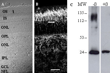
Figure 1. Rs1 Immunostaining and western blotting in adult pig retina
When incubated with sections of adult pig retina (bright field; A), anti-Rs1 antibody (B) demonstrated strong immunoreactivity within the interphotoreceptor matrix surrounding photoreceptor inner (IS) and outer segments (OS), as well as outlining cell bodies in the scleral region of the outer nuclear layer (ONL, arrow). There was equally prominent staining in the outer plexiform layer (OPL) and a subset of cells within the inner nuclear layer (INL), with staining percolating down through the inner plexiform layer (IPL) and visible in patches at the level of the ganglion cell (GCL) and nerve fiber (NFL) layers. Scale bar in B represents 30 μm. C: Western blots of retinal protein extracts prepared from adult pig retinas and run under non-reducing conditions (-β) demonstrated a prominent immunoreactive band about 25 kDa, and a high molecular weight band about 135 kDa. This latter disappeared in reducing conditions (+β) conditions, and represents a multimeric form. Molecular weight (MW) markers (in kDa) are shown to the left of the blots.