![]() Figure 3 of
Whikehart, Mol Vis 2005;
11:816-824.
Figure 3 of
Whikehart, Mol Vis 2005;
11:816-824.
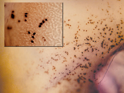
Figure 3. AP-BrdU staining of rabbit corneal endothelia at the point of wounding
BrdU staining at the periphery of a wounded rabbit corneal endothelium (the wound may be seen in the lower right corner of the large figure). The figure demonstrates that wounding induces cell division by taking up bromodeoxyuridine into new DNA. The image magnification is 27x; the magnification of the inset is 110x.