![]() Figure 2 of
Whikehart, Mol Vis 2005;
11:816-824.
Figure 2 of
Whikehart, Mol Vis 2005;
11:816-824.
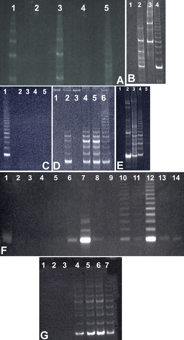
Figure 2. Telomerase activity in central, intermediate, and peripheral regions of human corneal endothelium
The figure demonstrates both the existence of telomerase activity in the peripheral endothelium (but not the central endothelium) and the elimination of contamination by Taq polymerase inhibitors in some tissue samples. The discovery of Taq polymerase inhibitors was initially a source of uncertainty in interpreting the results until its effects were eliminated by sample dilution. A: The initial demonstration of peripheral telomerase activity. Lane 1: HeLa cell control. Lane 2: Central-intermediate HCEC (16-year-old donor). Lane 3: Peripheral HCEC (16-year-old donor). Lane 4: Central-intermediate HCEC (66-year-old donor). Lane 5: Peripheral HCEC (66-year-old donor). B: This also demonstrates peripheral telomerase activity. Lane 1: Central-intermediate HCEC (55-year-old donor). Lane 2: Peripheral HCEC (55-year-old donor). Lane 3: TSR8 control. Lane 4: HeLa cell control. C: This panel, however, shows a complete lack of telomerase activity in peripheral endothelium as first seen in this study. Lane 1: HeLa cell control. Lane 2: Central-intermediate HCEC (26-year-old donor). Lane 3: Peripheral HCEC (26-year-old donor). Lane 4: Central-intermediate HCEC (55-year-old donor). Lane 5: Peripheral HCEC (55-year-old donor). D: Demonstrates the existence of a Taq polymerase inhibitor by mixing HeLa cells with HCEC cells. Lane 1: Peripheral HCEC alone (55-year-old donor from C). Lane 2: Peripheral HCEC mixed with HeLa cells. Lane 3: Buffer blank. Lanes 4,5: TSR8 controls. Lane 6: HeLa cells alone. E: Shows the existence of telomerase activity following dilution of a peripheral HCEC. Lane 1: Undiluted peripheral HCEC (23-year-old donor). Lane 2: Peripheral HCEC diluted 1:5 (same sample). Lane 3: Same diluted 1:25. Lane 4: HeLa cell control. Lane 5: Buffer blank. F: Demonstrates that extensive serial dilutions of central-intermediate and peripheral area HCECs only show telomerase activity in the endothelial peripheral area as taken from mixed aged donors (21-52 years). Lanes 1-5: Central-intermediate HCEC diluted 1:100, 1:400, 1:500, 1:600, and 1:800, respectively. Lanes 6-9: Peripheral HCEC diluted 1:75, 1:100, 1:200, and 1:400, respectively. Lanes 10-12: HeLa cells diluted 1:20, 1:40, and 1:1, respectively. Lanes 13,14: Buffer blanks. G: This panel gives no indication of central area (4 mm) telomerase activity following dilution of a 30-year-old donor. Lane 1: Buffer blank. Lane 2,3: Central HCEC undiluted and diluted 1:2, respectively. Lanes 4-6: Intermediate-peripheral HCEC undiluted, diluted 1:2, and diluted 1:10, respectively. Lane 7: HeLa cell control.