![]() Figure 1 of
Whikehart, Mol Vis 2005;
11:816-824.
Figure 1 of
Whikehart, Mol Vis 2005;
11:816-824.
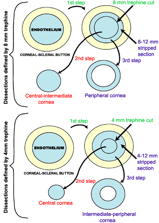
Figure 1. Flow diagram of method for dissecting corneal buttons for telomerase assays
Two types of dissections were performed: those divided by an 8 mm trephine generating central-intermediate (0-8 mm from center) and peripheral (8-12 mm from center) endothelial tissues and those divided by a 4 mm trephine generating central (0-4 mm from center) and intermediate-peripheral tissues (4-12 mm from center).