![]() Figure 6 of
Kirwan, Mol Vis 2005;
11:798-810.
Figure 6 of
Kirwan, Mol Vis 2005;
11:798-810.
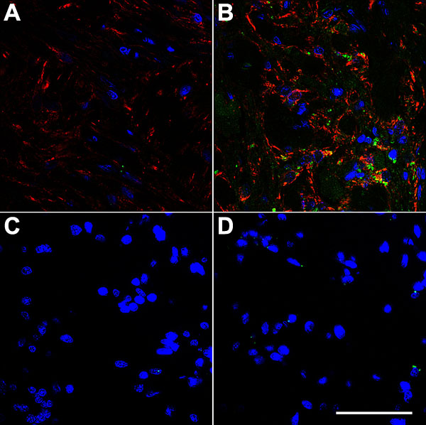
Figure 6. Double immunofluorescence histochemistry of normal and glaucomatous human optic nerve head tissue
Normal (A) and glaucomatous (B) optic nerve head sections were stained for EMMPRIN (green) and GFAP (red). EMMPRIN and GFAP staining were markedly increased in the glaucomatous sections compared to the normal controls. EMMPRIN was detected separate from and co-localized with GFAP (yellow) in the glaucomatous sections. Cell nuclei were DAPI stained (blue). No immunostaining was seen in the absence of primary antibody (C,D). The scale bar (D) represents 50 μm.