![]() Figure 1 of
Cameron, Mol Vis 2005;
11:775-791.
Figure 1 of
Cameron, Mol Vis 2005;
11:775-791.
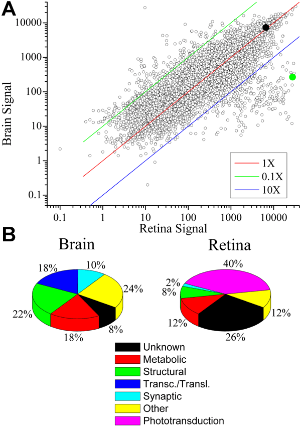
Figure 1. Gene expression profiles of adult zebrafish brain and retina
A: Scatter plot comparison of gene expression in control brain and retina. Each point represents a single transcript/spot on the array, plotted as a function of its signal amplitude (expression level) for brain and retina. The red line denotes equivalent signal amplitude for brain and retina, whereas the green and blue lines denote signal amplitudes that are 10X different from the equivalent signal amplitude. The filled green circle denotes rhodopsin, which is at much greater abundance in retina than brain, and the filled black circle denotes pgi1, which is approximately uniformly expressed (per total mRNA) in retina and brain. B: Pie chart summary of functional categories of the fifty most abundant transcripts of brain and retina (Table 10, Table 11).