![]() Figure 5 of
Seomun, Mol Vis 2005;
11:764-774.
Figure 5 of
Seomun, Mol Vis 2005;
11:764-774.
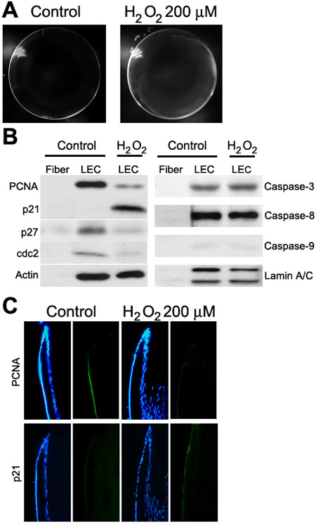
Figure 5. H2O2 induces lens opacification and the accumulation of p21Cip1 in rat lens
SD rat lenses were removed from eye and cultured on M199+0.1% BSA with or without 200 μM H2O2 for 1 day. A: Lens opacification was observed in the equatorial region spreading throughout the superficial cortex. B: Rat LECs and fiber cells were separately removed and analyzed using antibodies against the cell cycle (left) and apoptosis (right) related proteins. C: Rat lenses were embedded in paraffin, sectioned, and then immunostained with anti-PCNA (green, upper) and anti-p21Cip1 (green, lower). Nuclear stain with Hoechst (blue) was carried out to show the localization of LECs. The original magnification was x200.