![]() Figure 2 of
Seomun, Mol Vis 2005;
11:764-774.
Figure 2 of
Seomun, Mol Vis 2005;
11:764-774.
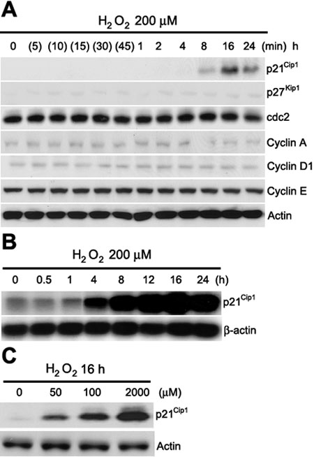
Figure 2. H2O2 induces the expression of p21Cip1
A: HLE B-3 cells were treated with 200 μM H2O2 at the indicated times. Cell lysates were prepared for western blot analysis. The expression of p21Cip1, p27Kip1, cdc2, cyclin A, cyclin D1, cyclin E, and actin were analyzed. B: Northern blot analysis for the induction of p21Cip1 by 200 μM H2O2 at indicated times. C: HLE B-3 cells were treated for 16 h in either the absence or presence various concentrations of H2O2 as indicated. The cell lysates were prepared and analyzed in the level of p21Cip1 by western blot. The data shown are representative of three independent experiments.