![]() Figure 1 of
Fan, Mol Vis 2005;
11:76-87.
Figure 1 of
Fan, Mol Vis 2005;
11:76-87.
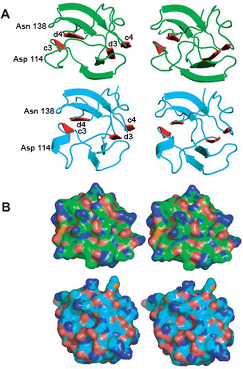
Figure 1. Comparison of rat γE-crystallin and human γD-crystallin C-terminal domains
Stereo representations comparing the C-terminal domain (III and IV motifs) of human γD-crystallin (green) and rat γE-crystallin (cyan). Both are views of the surface which is furthest away from the N-terminal domain. A: Ribbon representation, with motif III β-sheets at the bottom of the screen. The c and d β-strands from both motifs III and IV are colored red and labeled, as are the positions of Asp 114 and Asn 138 to orient the sequence. B: Surface renderings of the human γD-crystallin and rat γE-crystallin C-terminal domains, in the same orientation as in A. The carbon atoms are colored green or cyan as in A. Polar atoms are shown as follows; red for oxygen, blue for nitrogen, and orange for sulfur. There is a cleft (resembling a thumb print) in the surface of rat γE-crystallin running from the left side of the domain image to the middle. The dark blue protrusion to the top of this cleft is the side chain of Arg 163 (indicated with a white star) and the equivalent residue is also marked in the human γD-crystallin surface.