![]() Figure 3 of
Ma, Mol Vis 2005;
11:744-748.
Figure 3 of
Ma, Mol Vis 2005;
11:744-748.
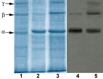
Figure 3. Experimentally induced high molecular crosslinks of vitreous collagen
SDS-PAGE and immunblotting of rabbit vitreous collagen were conducted. Coommassie-stained proteins were detected on lanes 1-3, and immunoblotting with anti-collagen antibodies is shown on lanes 4 and 5. Lane 1 contains bovine dermal type I collagen as molecular size markers. Lanes 2 and 4 contain rabbit vitreous collagen of a control (mock treated contralateral) eye, and lanes 3 and 5 contain rabbit vitreous collagen of the experimental eye. Arrows indicate the migration of α-, β-, and γ-components of vitreous collagen.