![]() Figure 2 of
Ma, Mol Vis 2005;
11:744-748.
Figure 2 of
Ma, Mol Vis 2005;
11:744-748.
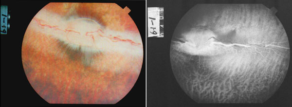
Figure 2. Experimentally induced retinal vein occlusion by photodynamic method
Fundus photography (left) and fundus fluorescein angiography (right) were conducted one day after photothrombosis. The retinal vein distal to the occlusion point became more tortuous, dark, and dilated which around with several surrounding small hemorrhages.