![]() Figure 2 of
Shan, Mol Vis 2005;
11:738-743.
Figure 2 of
Shan, Mol Vis 2005;
11:738-743.
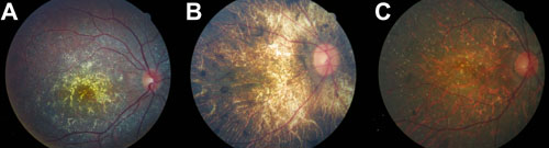
Figure 2. Fundus appearance of the patients with Bietti crystalline corneoretinal dystrophy
A: The fundus of the proband of family 27001 has many small yellowish-white sparkling spots distributed in the posterior pole with most of them located at the macular. B: The fundus of the proband of family 27002 has severe choroidal sclerosis with a few crystalline deposits. Pigmentation is observed in the peripheral area of the fundus. C: Patient 28029 has numerous yellow sparkling crystalline deposits evenly distributed in the posterior pole with pigmentation in the peripheral area.