![]() Figure 3 of
Seigel, Mol Vis 2005;
11:729-737.
Figure 3 of
Seigel, Mol Vis 2005;
11:729-737.
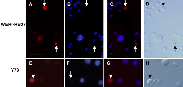
Figure 3. Co-localization of ALDH1 staining with Hoechst-dim cells in RB
For each cell type, we examined ALDH1 immunoreactivity along with Hoechst dye uptake in a dual-labeling experiment. Cancer stem cells would most likely be ALDH1-bright and Hoechst-dim. The top row (A-D) illustrates the same field of WERI-Rb27 cells. The bottom row (E-H) illustrates the same field of Y79 cells. A,E: ALDH1 immunoreactivity is shown in red. B,F: Hoechst dye uptake is shown in blue. C,G: Merged images of ALDH1 immunoreactivity and Hoechst dye uptake in WERI-Rb27 cells and Y79 cell line. D,H: Brightfield images. Arrows point to putative cancer stem cells that are immunoreactive for ALDH1 and Hoechst-dim. Scale bars represent 10 μm.