![]() Figure 2 of
Seigel, Mol Vis 2005;
11:729-737.
Figure 2 of
Seigel, Mol Vis 2005;
11:729-737.
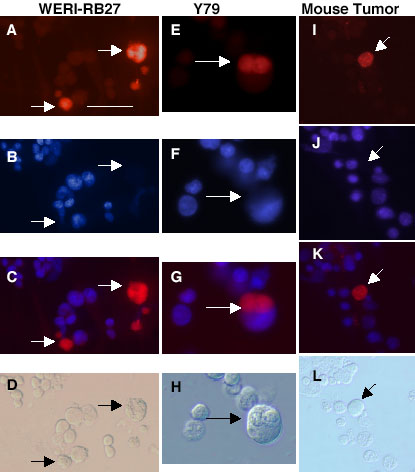
Figure 2. Co-localization of ABCG2 staining with Hoechst-dim cells in human and mouse RB
For each cell type, we examined ABCG2 immunoreactivity along with Hoechst dye uptake in a dual-labeling experiment. Stem cells that reside in a side population are typically ABCG2-bright and Hoechst-dim. The left column (A-D) illustrates the same field of WERI-Rb27 cells. The middle column (E-H) illustrates the same field of Y79 cells. The right column (I-L) shows the same field of mouse RB tumor cells. A,E,I: ABCG2 immumnoreactivity (red). B,F,J: Hoechst dye uptake (blue). C,G,K: A merged image (red and blue) of the top two rows. D,H,L: Brightfield images. Arrows point to bright red cells that are immunoreactive for ABCG2 and Hoechst-dim, characteristic of cancer stem cells. Scale bars represent 10 μm.