![]() Figure 4 of
Acosta, Mol Vis 2005;
11:717-728.
Figure 4 of
Acosta, Mol Vis 2005;
11:717-728.
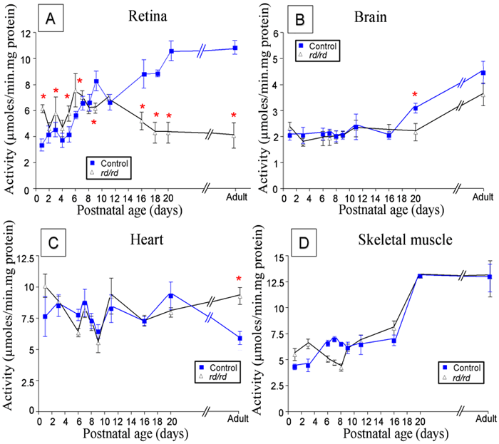
Figure 4. LDH activity in control and rd/rd tissues as a function of age
A: LDH activity in the control mouse retina steadily increased between P5 and P9 followed thereafter by a slow, gradual increment in activity until reaching adult levels. On the contrary, activity in the rd/rd retina was higher than control before P8 and decreased as photoreceptor cells died. Significantly different values are indicated by an asterisk (p<0.01). B: An increase in LDH activity was observed in the brain of both control and rd/rd mice after P16 with significantly different values observed at P20 (p<0.001). Total LDH activity was at all ages half the value found for the retina. C: LDH activity in the heart of both control and rd/rd mice was higher than the brain, despite having a similar LDH isoenzyme ratio. LDH activity did not vary through development although the adult value was slightly higher in the control mice (p<0.01). D: LDH activity in the skeletal muscle of control and rd/rd mice seemed to be similar to the retina. However, there were no differences between the rd/rd and control mousee LDH muscle activity.