![]() Figure 1 of
Acosta, Mol Vis 2005;
11:717-728.
Figure 1 of
Acosta, Mol Vis 2005;
11:717-728.
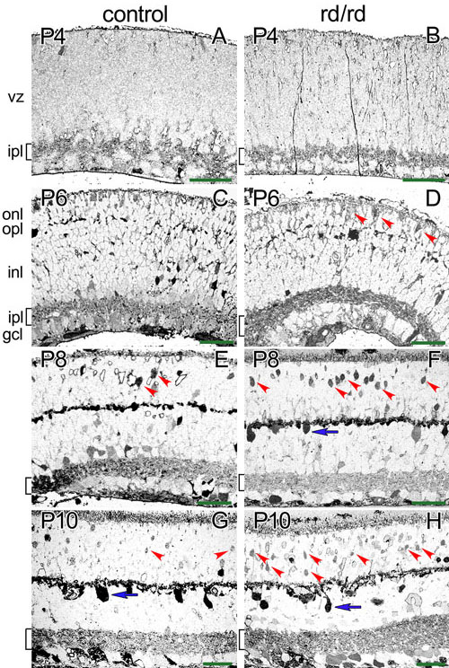
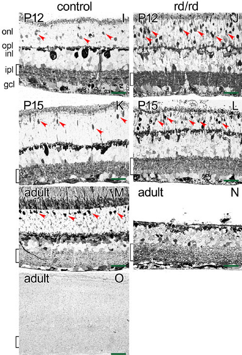
Figure 1. Control and rd/rd retinas immunolabelled for AGB
A,B: At P4, light AGB labeling was found in the developing inner plexiform layer. C,D: At P6, there was light labeling of the inner retina and few of the developing photoreceptor cells. In the rd/rd mouse, there was increased number of weakly labeled AGB photoreceptor cells. E,F: At P8, there was occasional labeling of horizontal cells (blue arrow), bipolar cells and the plexiform layers. At this age, an increased number of photoreceptors were strongly labeled in the rd/rd retina (red arrowheads). G,H: At P10, control and rd/rd retinas show labeling of horizontal cells (blue arrow) while in the rd/rd retinas there was a further increase in the number of labeled cells in the photoreceptor area. I,J: At P12, there was strong AGB labeling of the inner retina in both control and rd/rd retinas, while the labeling of the rd/rd photoreceptors continued to increase. K,L: At P15, the differences between control and rd/rd retinas were noticeable in the inner retina and photoreceptor area. M,N: In the adult, there was strong labeling of the inner retina in the rd/rd mice with no photoreceptor layer remaining. Panel O shows the absence of AGB labeling in an adult retina that was not incubated in the AGB solution. The different retinal layers are indicated in panels A, C, and I. The ventricular zone (VZ), inner plexiform layer (IPL), outer nuclear layer (ONL), outer plexiform layer (OPL), inner nuclear layer (INL), the inner plexiform layer (IPL), and the ganglion cell layer (GCL) are identified. The bracket on the side of each micrograph identifies the inner plexiform layer. The red arrowheads in panels D-M identify AGB labeled photoreceptors. The scale bar (green bar) corresponds to 20 μm.