![]() Figure 3 of
Chan, Mol Vis 2005;
11:697-704.
Figure 3 of
Chan, Mol Vis 2005;
11:697-704.
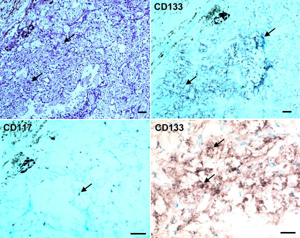
Figure 3. Stem cell markers in another hemangioblastoma
Positive cells (black bluish) for CD133 (arrows) highlight the VHL cells. Only a few positive CD117 cells (arrow) are seen. The upper left panel was stained with hematoxylin & eosin. The other panels represent avidin-biotin-complex counterstained with methyl green. The original magnifications of the upper and lower panels were 200x and 400x, respectively. The scale bars represent 50 μm.