![]() Figure 2 of
Chan, Mol Vis 2005;
11:697-704.
Figure 2 of
Chan, Mol Vis 2005;
11:697-704.
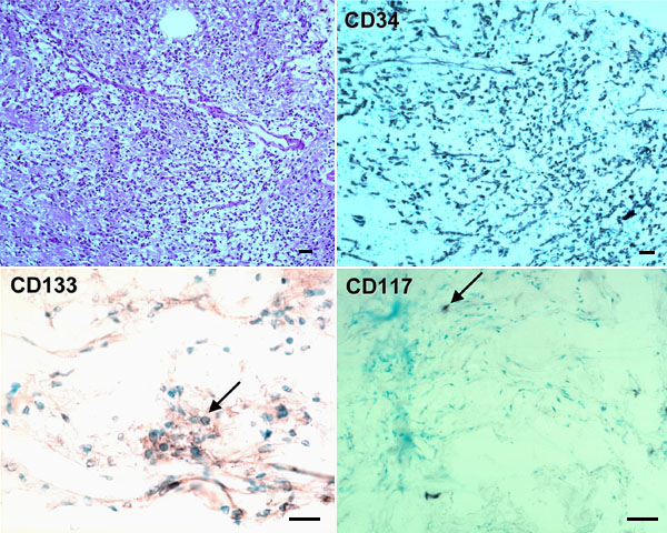
Figure 2. Stem cell markers in a retinal hemangioblastoma
Many positive cells (black bluish color) are stained with CD34 and CD133 (arrow). Only occasional positive CD117 cells (arrow) are seen. The upper left panel was stained with hematoxylin & eosin. The other panels represent avidin-biotin-complex counterstained with methyl green. The original magnifications of the upper and lower panels were 100x and 200x, respectively. The scale bars represent 40 μm.