![]() Figure 1 of
Chan, Mol Vis 2005;
11:697-704.
Figure 1 of
Chan, Mol Vis 2005;
11:697-704.
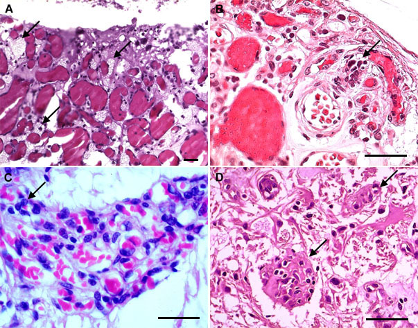
Figure 1. Tumorlet cells in ocular hemangiobalstoma
A: A classical VHL hemangioblastoma was composed of many vacuolated "stroma" cells (arrows) located between small vessels. B: Small tumorlet cells (arrow) were located adjacent to well-defined retinal vessels. C: Isolated small cells or tumorlet-like cells (arrow) were observed in a retinal hemangioblastoma. D: Several islands of tumorlets (arrows) are identified in an optic nerve hemangioblastoma. Sections were stained with hematoxylin & eosin; the original magnifications were 400x. The scale bars represent 50 μm.