![]() Figure 3 of
Delyfer, Mol Vis 2005;
11:677-687.
Figure 3 of
Delyfer, Mol Vis 2005;
11:677-687.
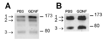
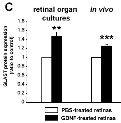
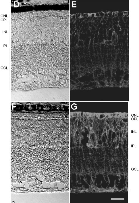
Figure 3. GLAST protein expression in rd1 mouse retinas in the presence and absence of GDNF
A,B: GLAST protein levels were studied in retinal organ cultures (A) and in vivo (B) in the presence and absence of GDNF. Western blots of total proteins (10 μg/lane) prepared from retinas 48 h after treatment with either GDNF or PBS, were probed with anti-GLAST. The blots demonstrated one band at about 76 kDa (GLAST monomeric form, labeled 3) and two bands at about 160 kDa (GLAST multimeric forms, labeled 1 and 2). C: GLAST protein was 1.48 times more abundant in GDNF-treated retinal organ cultures than in controls (6 samples per group). In vivo, GLAST protein was 1.25 times more abundant in GDNF-injected retinas than in controls (6 samples per group). Data are presented as the mean and SEM of the values obtained in three independent experiments (two asterisks indicate p<0.005, three asterisks indicate p<0.0005). D-G: Transverse sections of a PBS-injected eye and a GDNF-injected eye, from the same rd1 mouse, were examined with a confocal microscope. Sections were analyzed using the same parameters and exposure times. Pictures were always taken at the same distance from the optic nerve. D: PBS-injected eye, Nomarski optics. E: PBS-injected eye, immunostaining with anti-GLAST antibody. F: GDNF-injected eye, Nomarski optics. G: GDNF-injected eye, immunostaining with anti-GLAST antibody. Due to photoreceptor degeneration, the outer nuclear layer (ONL) is no longer composed of one or two layers of photoreceptors. Pictures are representative of four independent experiments with similar results. The outer plexiform layer (OPL), inner nuclear layer (INL), inner plexiform layer (IPL), and ganglion cell layer (GCL) are also labeled. The scale bar represents 20 μm.